Percutaneous Nephrolithotomy
Home / Procedure Detail

Percutaneous Nephrolithotomy
- Procedure
- Percutaneous Nephrolithotomy
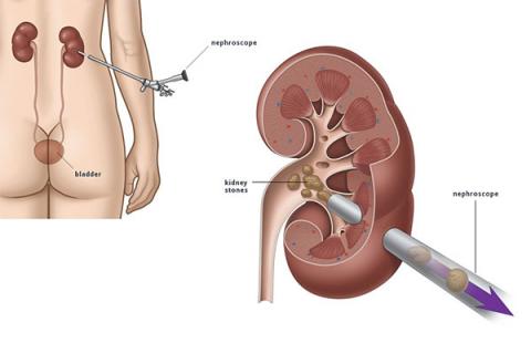
A percutaneous nephrolithotomy is an option for removing kidney stones from the body when they can't pass on their own. “Percutaneous” means that it takes place through the skin. A surgeon creates a passage between the skin on the back and the kidney using special instruments that travel via a tiny tube in your back. Stones are located and removed from your kidney by means of special instruments passed through a tiny tube in your back.
Percutaneous nephrolithotomy is most often used to treat larger stones or when less-invasive procedures fail to work or are unavailable.
Why it's done
Percutaneous nephrolithotomy is a medical procedure used to remove large kidney stones. This surgery is typically recommended in the following cases:- The patient has large kidney stones that are blocking more than one branch of the collecting system of the kidney, known as staghorn kidney stones.
- The patient's Kidney Stones are larger than 0.8 inches (2 centimeters) in diameter
- Large Stones are present in the tube connecting a patient's kidneys and bladder (ureter). -Other therapies have failed

Procedure Primary Points
- Pre-procedure: General anesthesia is administered in the hospital.
- Procedure: Needle inserted into kidney calyx for access.
- Surgeon guides needle placement using imaging (X-ray, CT, ultrasound).
- Stones are removed using specialized tools through a sheath; nephrostomy tube may be inserted.
- Post-procedure: Hospital stay, physical activity restrictions, follow-up visit, stone analysis, preventive measures.
What You Can Expect
Before the procedure
Percutaneous nephrolithotomy is usually done under general anesthesia in the hospital. You won't be awake for the surgery and you won't feel any pain because of general anesthesia. The first step of the procedure, on rare occasions, may be completed in radiology. Then, after you've been moved to surgery, you'll receive general anesthesia.During the procedure
A needle is inserted into the urine-collecting chamber of the kidney (calyx) to start. The path of this needle becomes the access for performing the procedure. A surgeon or trained radiologist guides the needle's placement through X-ray, CT, or ultrasound images. This can happen either in the operating room or the radiology department. You may have a flexible tube (catheter) passed into your kidneys from the bladder and urethra. The urinary tract starts with the urethra, which is a tube that urine leaves through. The connection between a kidney and a bladder is the ureter. A tiny camera inserted via this catheter allows your doctor to see the needle as it's inserted into the kidney and other operations during surgery. Following this, the surgeon inserts a tube (sheath) along the needle's route. The surgeon fractures and removes the stones using specialized tools that pass through the sheath. A nephrostomy tube may then be inserted in this same passageway. During recovery, a patient can wear a bag containing urine outside of his or her body via this tube. If more kidney stones or fragments are required to be removed during the healing period, this tube allows for additional access to the kidney. The kidney stones are sent to a lab to check what types of stones they are. Knowing what type of kidney stones you have may help your care provider suggest ways to prevent future stones.After the procedure
After the operation, you might be in the hospital for 1 to 2 days. For 2 to 4 weeks after surgery, you may need to avoid physical activity that puts strain on your incision. After approximately a week, you should be able to return to work. If you have kidney drainage tubes after surgery, be on the lookout for bleeding. If you detect blood or large clumps of ketchup-like blood in your urine or drain, go to the emergency room. If you have a fever or chills, see your primary care doctor or the surgical team. These might be indications of an infection, and you may require urgent care. If you're experiencing severe pain that doesn't respond to pain medication, call your doctor.Results
After surgery, you'll get your surgeon or primary care provider 4 to 6 weeks later for a check-up visit. If you have a nephrostomy tube for drainage, you could go home sooner. You may undergo an ultrasound, X-ray, or CT scan to inspect for any stones that may remain and ensure that urine is passing properly from the kidney. Your surgeon will remove the nephrostomy tube after administering a local anesthetic if you have one. Your surgeon or main care physician might advise blood tests to discover what caused the kidney stones. You can also discuss ways to avoid getting more kidney stones in the future.Medical Procedures
MedEx did help me a lot not only for connecting with clinics but also for other miscellaneous items such as visa extention. I am really sastify with the services received from MedEx. I've recommended to some of my friends to connect with MedEx too if they have plan to go for medical trip to BKK. 🙂
Engyin HtunSingapore 
Highly recommend to MedEx .They are professional and amazing team.
MedEx Staffs should have closed relationships with hospitals staffs to get more information and services.So,they can give the best services to patients.
Thomas FlyerMyanmar 
Owing to my heart pacemaker implant case, if I got a chance to refer anyone, without blinking my eyes I would refer to Vejthani Hospital. I am very grateful.
Bhagwan Ratna TuladharKathmandu, Nepal 
MedEx was very kind, patient, supportive and very helpful with my needs for seeing doctors and doing physical therapy here in Thailand. MedEx fulfilled everything I needed with my stay here with prompt actions, and I highly recommend MedEx if you are coming to Bangkok for medical treatment.
Ko Sai Aung Lwin TunMyanmar 
Previous
Next


